Description
Modern ophthalmology is rapidly advancing with technology that allows eye disease to be detected earlier and treated more precisely than ever before.
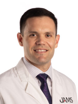
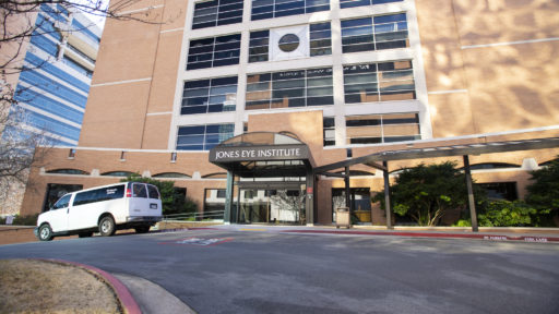
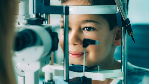
In this video, Dr. Ryan Wood of the UAMS Health Jones Eye Institute explains how optical coherence tomography (OCT) imaging allows specialists to see detailed structures inside the eye at a microscopic level. This technology helps identify eye conditions before symptoms appear, allowing for earlier intervention and better long term outcomes.
Dr. Wood also discusses the future of eye care, including promising developments in stem cell therapy and 3D printed tissue engineering. These innovations may one day reduce the need for donor corneas and improve compatibility for patients requiring eye tissue replacement.
At the UAMS Health Jones Eye Institute, advanced diagnostic tools and ongoing research are helping transform the way vision problems are diagnosed, treated, and managed.
Video Player

Transcript
One of the reasons I came to the Jones Eye Institute is because of their technology. They have lots of instruments to use, not only to diagnose ocular problems, but to treat those problems. One of my favorites is the OCT. The OCT isn’t necessarily new equipment, but it’s been updated over the years, and it helps measure problems inside the eye down to the micron. So we can observe those problems and often detect them before patients know about them. We can also treat them before they cause any vision loss the patient might otherwise experience in the future.
Exciting things in the future for me would be centered around stem cell treatment. Right now, cornea conditions often have to be replaced like an organ, with donor tissue. Whereas now, researchers are working not only on using stem cells to treat those problems, but on creating tissues with a 3D printer from those cells, to make tissues that are more compatible with a person’s own body, as opposed to risking rejection like a typical donation.