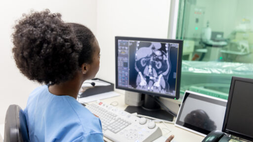
A VQ scan (Ventilation-Perfusion Scan) is a medical imaging test that evaluates the airflow (ventilation) and blood flow (perfusion) in the lungs. It’s commonly used to diagnose or rule out a pulmonary embolism (PE), which is a blood clot in the lungs, but can also assess other lung conditions.
Components of a VQ Scan:
- Ventilation Scan:
- This part of the test assesses how well air is reaching all parts of the lungs.
- The patient inhales a gas (often xenon or technetium-labeled aerosol) that contains a small amount of radioactive material.
- A gamma camera takes images of the lungs as the gas is inhaled, showing how air moves through the lung tissue.
- Perfusion Scan:
- This part measures the blood flow in the lungs.
- A different radioactive tracer (usually technetium-99m) is injected into a vein, which travels to the lungs.
- The gamma camera then takes images of the lungs, highlighting areas with good or poor blood flow.

