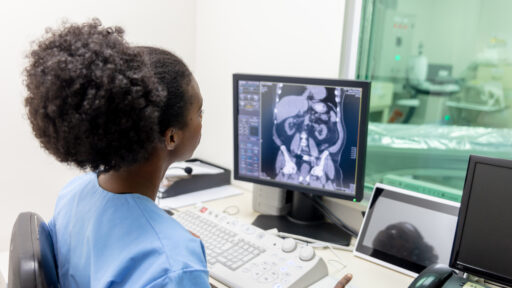
A Cisternography, also known as a Cisternogram, is a type of nuclear medicine imaging test used to evaluate the flow of cerebrospinal fluid (CSF) within the brain and spinal cord. This test is particularly useful for diagnosing conditions related to CSF circulation, such as cerebrospinal fluid leaks, hydrocephalus, or normal pressure hydrocephalus (NPH). Here’s how it works and what it involves:
How It Works:
- Radiotracer Injection: The procedure begins with the injection of a small amount of a radioactive tracer into the cerebrospinal fluid. This is typically done through a lumbar puncture (spinal tap), where the tracer is introduced into the subarachnoid space around the spinal cord.
- Tracer Distribution: Once injected, the tracer mixes with the cerebrospinal fluid and begins to circulate throughout the brain and spinal cord. The movement of the tracer through the CSF pathways is tracked over time.
- Imaging: After the tracer is administered, a series of images are taken using a gamma camera at various intervals (typically over several hours or days) to monitor the flow and distribution of the cerebrospinal fluid. The images can reveal any abnormalities in the circulation or absorption of CSF.
- Image Analysis: The collected images are analyzed to determine if there are any blockages, leaks, or other disruptions in the CSF pathways. For example, in cases of suspected CSF leak, the scan can help identify the exact location of the leak.

