PT Program Turns to Plasticized Remains to Teach Anatomy
| July 14, 2017 | John Jefferson, Ph.D., P.T., chair and associate professor of the Department of Physical Therapy (PT) in the College of Health Professions, thought long and hard about the best way to teach anatomy to physical therapy students when he was setting up the program.
Many PT programs operate a typical anatomy lab in which the students dissect the cadavers so they can study human anatomy. Jefferson considers the whole dissection process a waste of time for PT students since they don’t need those particular skills.
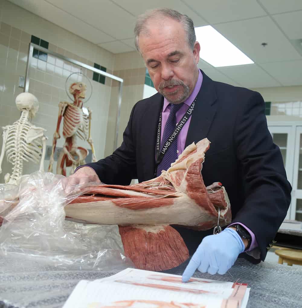
“PT students don’t need to practice cutting since they’re not going to be performing surgery,” he said. “What they do need is to see where everything is in 3D.”
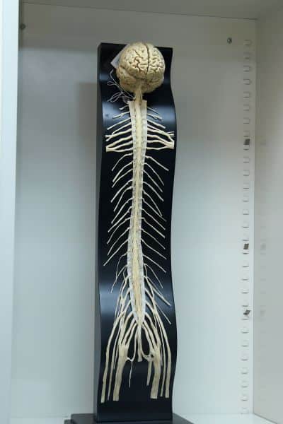
This cadaver shows the brain, spinal cord and nerves that make up the central nervous system.
He considered sharing cadavers with the medical school, which would let the medical students complete the dissections and then the PT students could study the muscles and tendons. But that would have required shipping the bodies from Little Rock to Fayetteville and then shipping them back at the end of the semester for proper disposal.
It also would have locked the PT program into a one-semester anatomy course, which Jefferson thinks is a problematic way to teach students about the subject.
So Jefferson looked for an out-of-the-box solution.
He investigated plasticized cadavers, which are bodies that have been through a multi-step process that embalms the specimens to prevent decomposition; dissects them to the desired depth; places them in a solvent bath that removes body water and soluble fats; and puts them in a vacuum where the solvent is extracted and replaced by liquid plastic that is then cured (hardened) while the body or body part is held in position by wires, needles, clamps and foam blocks. Dissection and plastination of an entire body requires about 1,500 working hours and normally takes about one full year to complete.
After working with a German company that prepares and sells plasticized cadavers for medical schools, UAMS purchased nearly $225,000 worth of plasticized remains for the PT program’s anatomy lab.
“I chose the company we worked with because of their excellent reputation and the quality of their work,” said Jefferson. “The company is also highly ethical. They only use remains of people who willingly donated their bodies for the expressed purpose of plastination and only sell their products to qualified users, such as medical schools, hospitals and museums.”
Two years later and the program is ordering more specimens for the lab, thanks to nearly $150,000 in additional funding provided by the UAMS provost’s office.
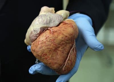
John Jefferson, Ph.D., P.T., holds a plasticized human heart.
“At the time we started, we got the bare minimum of what we needed for the lab, including two whole bodies and one arm and leg that we use a lot because of the nature of physical therapy,” said Jefferson. “It certainly can get pretty cramped when you crowd 24 students around two or three limbs, so we are grateful that the provost has provided us with additional funds for our lab.”
While the initial costs for the lab were sizable, it will be less expensive in the long run than operating a typical anatomy lab, which costs approximately $30,000 a year, Jefferson said.
One of the things that Jefferson likes best about using plasticized remains is the flexibility they offer him in teaching anatomy to the students.
“Medical students have to learn the anatomy of the entire body in one semester. Every muscle, every nerve, every joint, every organ — they have to try and learn it all in one semester,” he said, adding that this grueling schedule is dictated by the use of cadavers, which can only be used for a limited time.
“But the plasticized remains, which we hope will last for 20 or more years, allow us to spread our anatomy curriculum into five separate courses taught over four semesters,” he said.
PT students have specialized anatomy courses in the brain and nervous system, heart and lungs, upper limb, lower limb and the spine.
“The plasticized remains allow our students to see in 3D what they see in 2D in the anatomy book,” he said. “I just can’t stress how important a learning tool these specimens are for our students.”
“We are only on our second class of PT students and I’m already seeing an improvement in how these students are learning anatomy versus those I taught using the dissection method years ago,” he said.